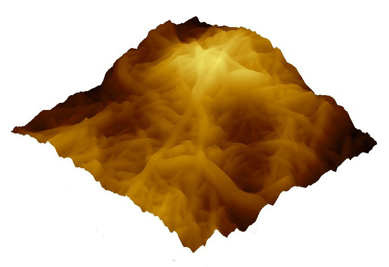
The surface structure of a material plays an important role in endothelialization of a substrate. Here the surface of a nanofibrous membrane was characterized by atomic force microscope.
Oddný Björgvinsdóttir
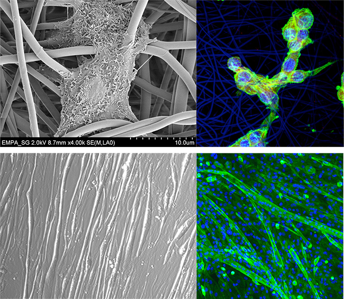
Figure 1. Initial studies for verification of material system biocompatibility. C2C12 myoblasts seeded on electrospun EVOH scaffold. SEM image of single cell attached to the fiber network on the left and CLSM image of cells 24 hours after the seeding process on the right. C2C12 cells electrosprayed with 15 kV onto TCPS after 7 days in differentiation medium. Cells are viable after the process and no changes in phenotype could be observed. The characteristic differentiation into myotubes was analysed via optical microscopy and CLSM.
Lukas Weidenbacher
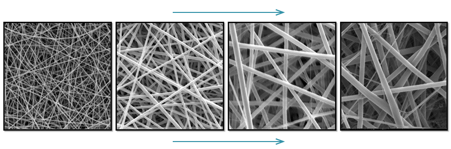
Figure 2. Cospun PCL and gelatine fibers. Increasing the polymer concentration in the spinning solution gives fibers with increasing fiber thickness.
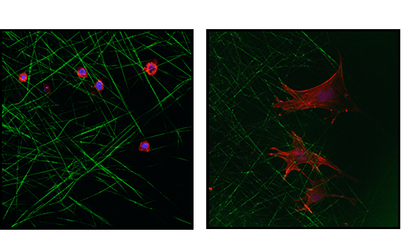
Figure 3. HUVECs after 16 hours in culture on electrospun fibers.
a) HUVECS on PCL fibers b) HUVECs on gelatin coated PCL fibers.
Oddný Björgvinsdóttir